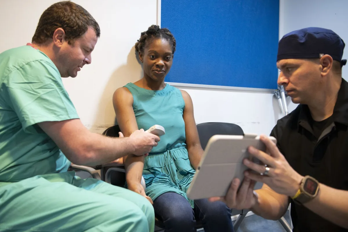
Ultrasound Spurs Bone Regeneration Through Molecular Pathways
Researchers are uncovering the intricate molecular mechanisms by which low-intensity pulsed ultrasound (LIPUS) promotes bone regeneration, paving the way for enhanced therapeutic strategies. A team from Capital Medical University in Beijing, China, has identified a specific signaling pathway, termed CaN/NFATC3/Id3/E2A/Bmp4, that explains how LIPUS stimulates the body's natural bone-healing processes. Their findings, published in the International Journal of Oral Science on September 14, 2026, pinpoint Id3 as a molecular 'brake' that typically limits ultrasound-induced bone formation. Following ultrasound stimulation, Id3 levels were significantly reduced, suggesting a key target for intervention.
Building on this discovery, the researchers developed an Id3 inhibitory peptide, known as rID3. When administered in conjunction with LIPUS, this peptide demonstrated an enhanced ability to promote osteogenesis and accelerate bone defect healing in mouse models. This dual approach, combining LIPUS with targeted molecular inhibition, represents a promising non-invasive strategy for improving bone regeneration. Professor Songlin Wang and Associate Professor Lei Hu, the corresponding authors of the study, highlighted the potential convenience and lack of systemic side effects compared to traditional drug-based treatments, suggesting a significant advancement in non-invasive bone healing techniques.
The broader field of sonogenetics, which merges ultrasound with genetic engineering, is also showing considerable promise for bone repair. LIPUS itself acts as a non-invasive mechanical stimulus, capable of activating cellular signaling pathways crucial for bone formation. However, the current therapeutic efficacy of LIPUS in repairing bone defects is often suboptimal, underscoring the critical need to identify molecular targets that can amplify its regenerative effects. The work from Capital Medical University directly addresses this gap by elucidating a fundamental molecular pathway.
New Ultrasound Platform Targets Early Stress Fracture Detection
In parallel advancements, researchers at the Johns Hopkins Applied Physics Laboratory (APL) in Laurel, Maryland, are developing a novel ultrasound-based platform designed to detect stress fractures in their earliest stages. This technology, named Osteo Surface Echo Evaluation (OsteoSEE), utilizes surface acoustic waves to identify subtle microstructural changes and nascent fractures within the dense outer layer of bone, known as cortical bone. A significant advantage of OsteoSEE is its non-invasive nature, completely avoiding the use of ionizing radiation associated with conventional X-ray imaging.
The OsteoSEE platform was recognized as a 2026 R&D 100 Award winner, acknowledging its innovative approach to early fracture detection. Anna Knight, the principal investigator for OsteoSEE and a senior biomechanical engineer at APL, emphasized the technology's critical application in supporting military readiness. She noted that fully developed stress fractures can incapacitate individual service members, and the widespread occurrence of this overuse injury diminishes overall mission readiness across the force. Early detection through OsteoSEE could enable timely intervention, preventing more severe injuries and maintaining operational capability.
OsteoSEE addresses limitations inherent in current diagnostic methods. Traditional X-rays often lack the sensitivity to pick up early-stage or hairline stress fractures. While advanced imaging techniques like MRI offer greater detail, they are considerably more expensive and require access to specialized, often limited, facilities. OsteoSEE circumvents these issues by employing commercially available ultrasound transducers. These transducers direct sound waves towards the bone at precise angles and frequencies, allowing for detailed analysis of the bone's surface and subsurface structure without the drawbacks of radiation exposure, high cost, or facility dependence.
Ultrasound Enhances Detection of Hidden Bone Infections
Beyond fracture healing and detection, ultrasound technology is also proving invaluable in identifying complications associated with bone injuries, specifically fracture-related infections (FRI). A study conducted at Erasmus Medical Center in Rotterdam suggests that a technique adapted from prosthetic joint surgery can significantly improve the detection of bacteria on fracture-fixation hardware, such as plates, screws, and nails. By subjecting removed hardware to ultrasound waves and culturing the fluid dislodged from the implant surface, researchers found they could identify bacteria that standard tissue cultures often miss.
This improved diagnostic capability has a direct impact on patient care, leading to changes in diagnoses and antibiotic treatment decisions for a meaningful proportion of patients. Fracture-related infection is a serious complication of operative fracture care, with reported infection rates varying widely depending on the fracture type. Open fractures, where the bone pierces the skin, present a direct pathway for bacteria and have higher infection rates, potentially reaching up to 29 percent. Diagnosing FRI is notoriously challenging due to variable symptoms and the frequent failure of conventional culture methods, which can yield negative or inconclusive results. Literature indicates that a significant percentage of confirmed FRI cases show no bacterial growth on standard cultures, forcing surgeons to treat empirically.
The effectiveness of ultrasound sonication in this context stems from its ability to overcome the problem of biofilm. Bacteria residing on implanted metal do not exist as free-floating organisms; instead, they form a protective, sticky matrix that adheres tenaciously to the implant surface. Ultrasound waves create cavitation—the rapid formation and collapse of microscopic bubbles—which can effectively disrupt this biofilm and release the bacteria or their components into the surrounding fluid, making them detectable by subsequent culturing.
Ultrasound Aids Sustainable Pharmaceutical Compound Synthesis
In a different application of acoustic technology, researchers are utilizing ultrasound to facilitate the sustainable production of valuable molecules relevant to pharmaceutical research. Studies from the IPC PAS (Institute of Physical Chemistry of the Polish Academy of Sciences) demonstrate that lignin, a highly abundant natural polymer and a major byproduct of wood processing, can be transformed into useful compounds using light and ultrasound. This approach offers a cleaner alternative to traditional synthesis methods that often rely on fossil feedstocks, high temperatures, strong oxidants, and potentially costly or difficult-to-purify metals.
The process involves using light to activate a catalyst, while ultrasound waves generate cavitation. This cavitation creates localized energy hotspots precisely where the chemical reaction occurs, avoiding the need to heat the entire system to extreme temperatures. Dr. Marta Paszkiewicz-Gawron and her colleagues successfully converted coniferyl alcohol into dehydrodiconiferyl alcohol (DHCA), a compound formed by linking two coniferyl alcohol units. Under optimized conditions, this reaction achieved 100% substrate conversion and 54% selectivity for DHCA, remaining stable over a 24-hour test period. Acoustic field mapping revealed that ultrasound's role extends beyond simple mixing; its intensity and distribution can actively influence and direct the types of products formed.
What This Means
The recent developments in ultrasound technology signify a multi-faceted leap forward in biomedical applications. For bone healing, the identification of specific molecular pathways like CaN/NFATC3/Id3/E2A/Bmp4 and the development of targeted peptides like rID3 offer the potential for significantly enhanced, non-invasive regenerative therapies. This could translate to faster recovery times and improved outcomes for patients with bone defects, moving beyond the limitations of current treatments by directly influencing cellular mechanisms. The application of ultrasound in detecting hidden bone infections also promises more accurate diagnoses and tailored antibiotic strategies, reducing the risks associated with undiagnosed or mistreated FRI.
In the realm of diagnostics, the OsteoSEE platform represents a critical advancement for early stress fracture detection, particularly for high-risk populations such as military personnel. By providing a non-ionizing, cost-effective, and accessible method for identifying microstructural bone damage before it becomes clinically apparent, this technology could revolutionize injury prevention and management, thereby enhancing readiness and reducing long-term disability. Furthermore, the use of ultrasound in sustainable chemical synthesis highlights its versatility, offering greener and more efficient routes to producing valuable compounds for pharmaceutical research, reducing environmental impact and production costs.
Looking ahead, further research will likely focus on refining these ultrasound-based techniques. For bone regeneration, this may involve optimizing peptide delivery and LIPUS parameters for human clinical trials. In fracture detection, the scalability and real-world deployment of OsteoSEE will be key. The continued exploration of ultrasound's role in disrupting biofilms and its application in synthesizing complex molecules also suggests a broad and expanding impact for this technology across various scientific and medical fields.




💬 Comments (0)